Facial Paralysis Reconstruction After Blood Cancer Treatment
A multi-technique approach to restore facial function and smile.
At a Glance
The Challenge
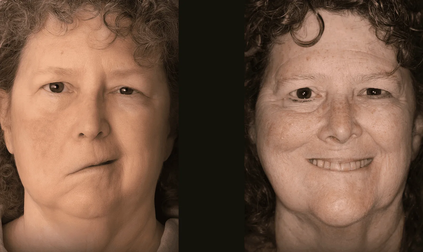
This patient experienced facial paralysis as a result of a blood cancer and its treatment. The loss of facial nerve function left them unable to smile or move the affected side of the face, creating functional difficulties and significant emotional distress during an already challenging recovery.
The Solution
Dr. Jowett used a multi-vector reconstruction. Healthy nerves that normally contribute to shoulder movement and chewing were rerouted to supply facial movement, and a small segment of gracilis muscle from the thigh was transplanted to rebuild a smile mechanism. The combination was selected to address both resting tone and intentional smile movement in this patient.
Technical Insight
Procedure: Spinal accessory nerve transfer, masseteric nerve transfer, and free gracilis muscle transfer.
Surgical Note: This case illustrates a comprehensive, multi-modal approach for severe or long-standing paralysis. Nerve transfers (spinal accessory, masseteric) provide axonal input to native musculature where viable. The free gracilis flap adds new contractile muscle tissue to restore dynamic excursion of the oral commissure (smile) when native muscles have atrophied or require augmentation.
First Stage Surgery: Multiple Nerve Transfers
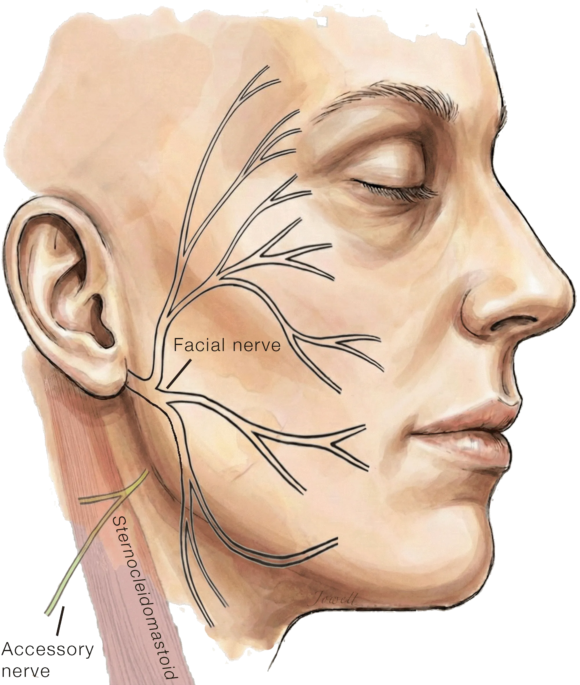
Accessory to Facial Nerve Transfer for Facial Reanimation
In facial paralysis resulting from injury to the facial nerve near the brainstem, the nerve no longer delivers electrical signals to the muscles of expression. However, the nerve branches within the face itself remain otherwise intact, ready to be reconnected to new power sources to restore tone and movement.
Common donors include hypoglossal and masseteric branches. In a patient who already has difficulty swallowing or chewing, an accessory-nerve branch may be considered instead. The accessory nerve supplies neck and shoulder muscles; using it as a donor requires discussion of possible shoulder or neck weakness along with the intended facial benefit.
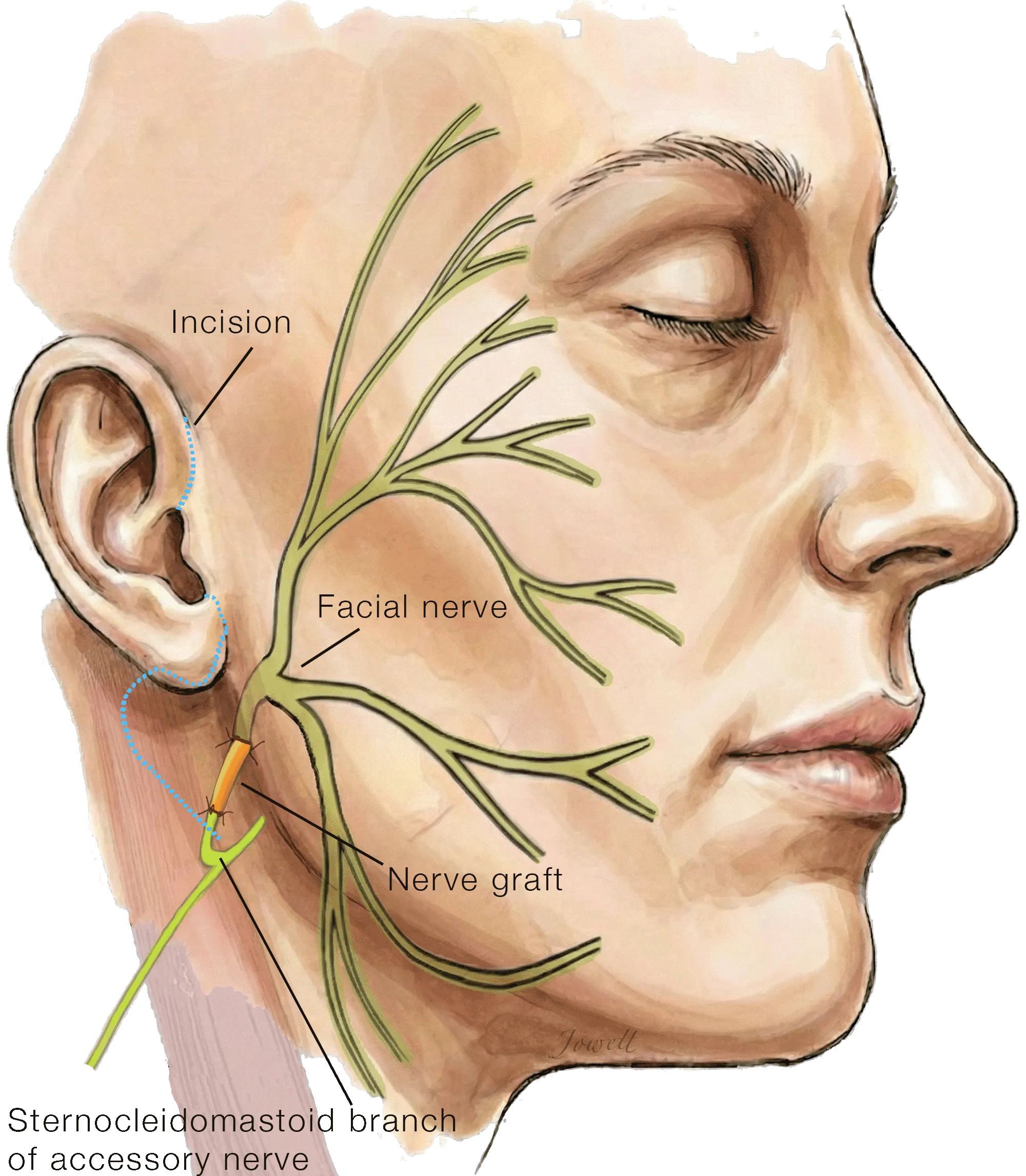
Accessory to Facial Nerve Transfer for Facial Reanimation
Through a small incision, a branch of the accessory nerve carrying fibers that supply the sternocleidomastoid muscle in the neck are rerouted, using a small nerve graft, to the muscles of facial expression to improve facial tone and symmetry without impacting shoulder function.
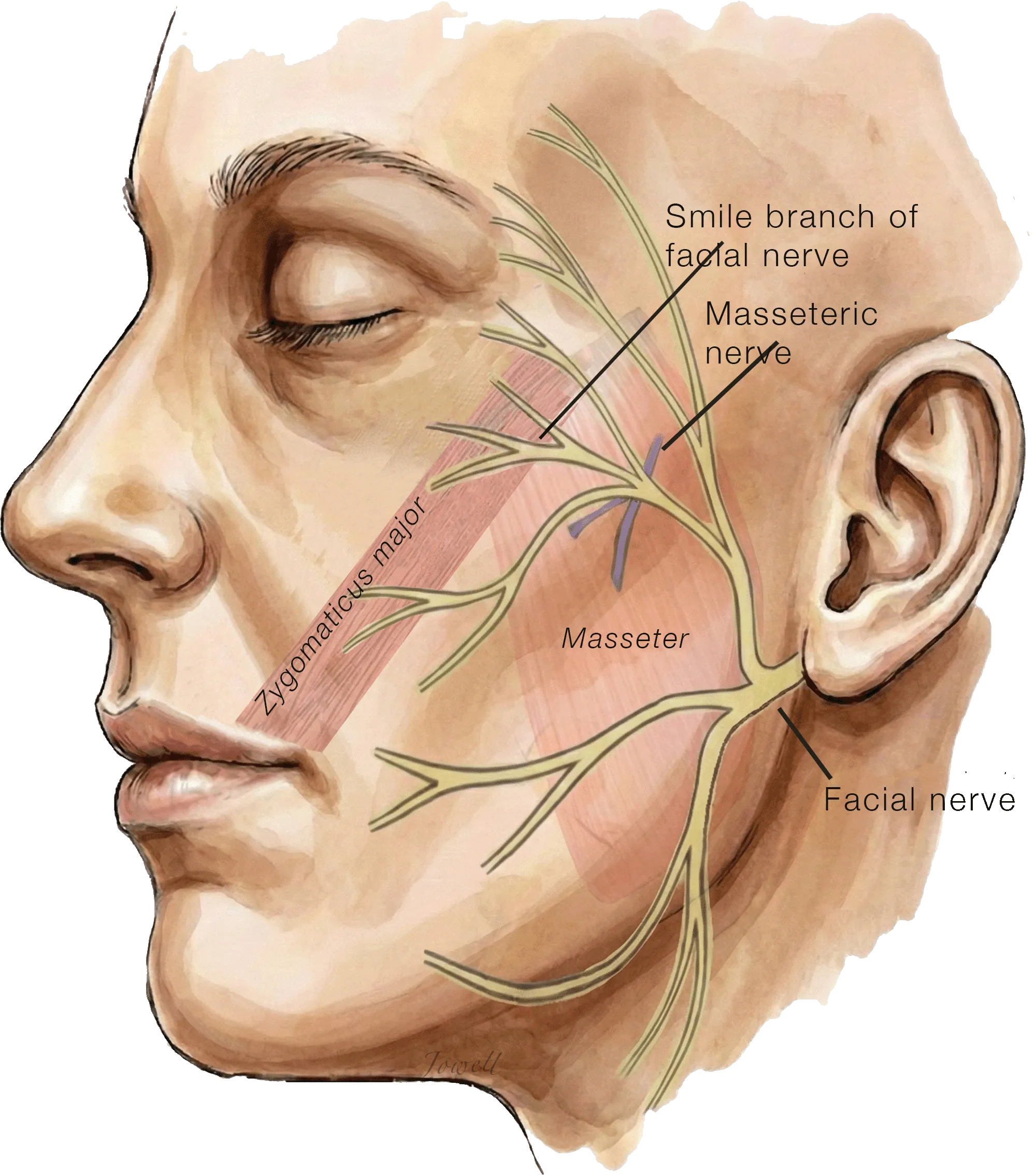
Cross-Facial Nerve Grafting "Donor" Nerves
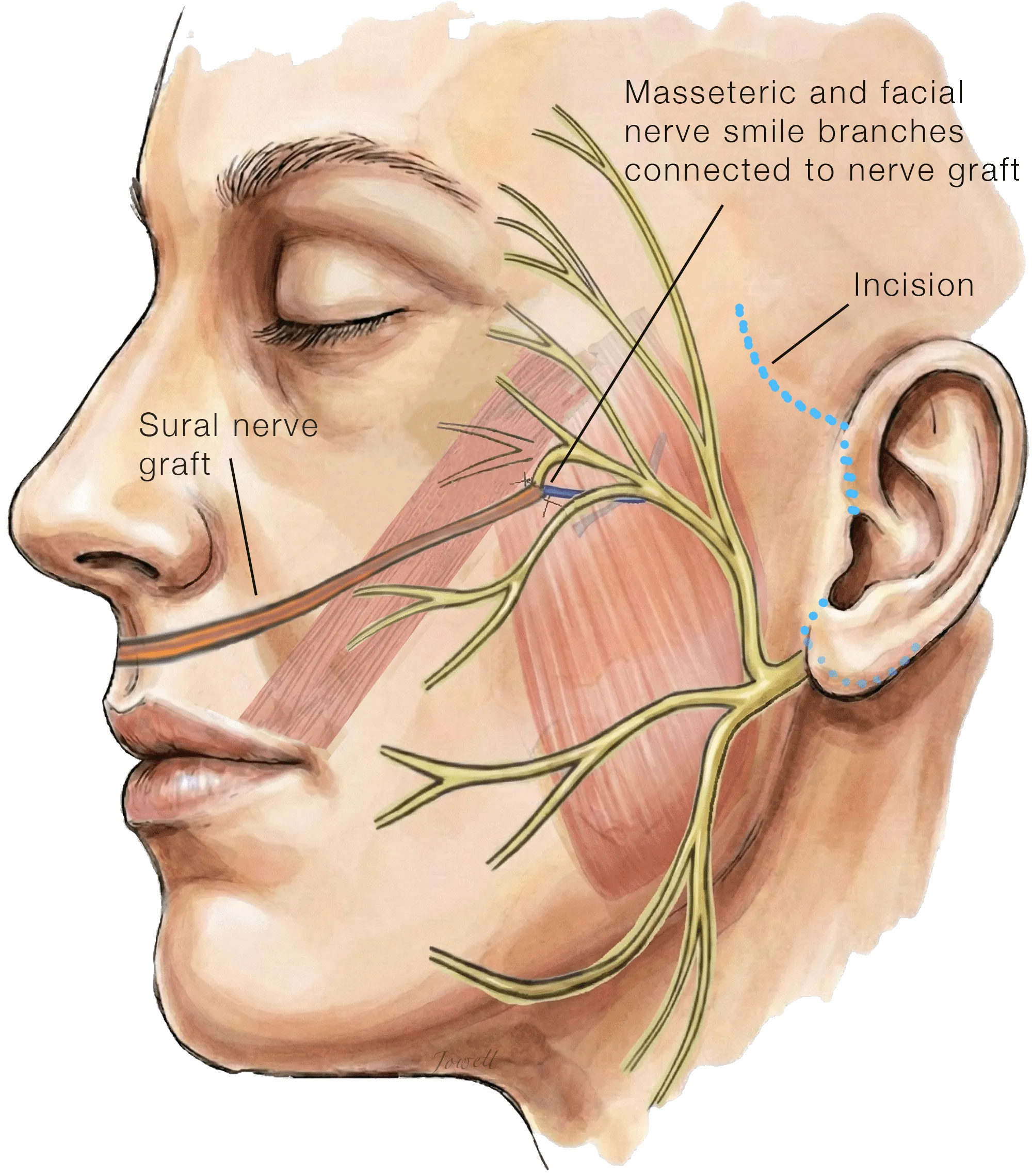
This diagram illustrates the anatomy on the unaffected (healthy) side of the face. To restore a smile on the paralyzed side, we identify two distinct 'power sources' on this healthy side:
Smile nerve: A redundant branch of the facial nerve connected to the smile muscle (Zygomaticus major).
Chewing nerve: A branch of the nerve used for chewing (masseter nerve).
After surgically locating these specific distal branches, we prepare to connect them to a nerve graft bridge that will carry their signals across the face to power smile movements on the paralyzed side.
Masseteric-Supercharged Cross-Facial Nerve Graft
The long nerve graft harvested from the lower leg is tunneled under the skin across the upper lip via a facelift incision and connected to the two 'power sources': the smile branch and the chewing branch. This is a masseteric-supercharged cross-facial nerve graft, not dual innervation of the muscle itself: both signals travel in one graft and later reach the transplanted muscle together.
Over the next several months, nerve fibers from the healthy side will grow across the nerve graft, where they will later power muscle transferred from the thigh to the face. The smile-nerve fibers keep the timing of a spontaneous smile; the chewing-nerve fibers add power.
Second Stage Surgery: Functional Muscle Transfer for Smile Reanimation
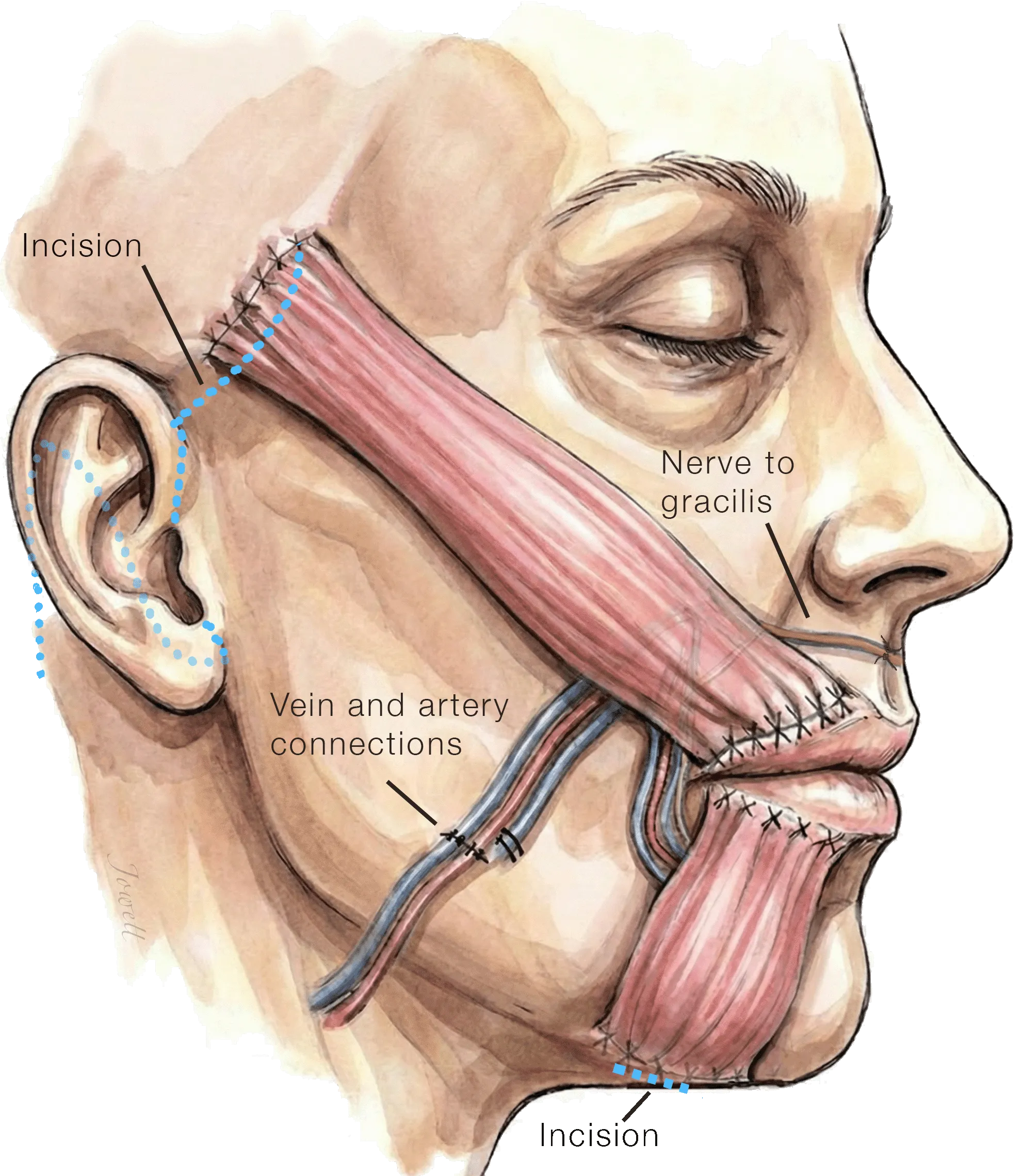
Dual-Vector Free Gracilis Muscle Transfer for Smile Reanimation Neurotized by Cross-Facial Nerve Graft
After a delay of several months to allow nerve fibers to grow across the nerve graft, a small strip of gracilis muscle is taken from the thigh, divided into two parts, and carefully placed into the face to rebuild the smile. This is done through a well-camouflaged facelift incision, along with small incisions inside the mouth and under the chin.
The muscle’s artery and vein are then connected to blood vessels in the face, and its nerve is connected to the cross-facial nerve graft that was placed during the first stage of surgery.
The Outcome
One year after the final stage of reconstruction, the patient has achieved a remarkable restoration of facial symmetry and expression. The smile is broad and natural, and the overall facial tone is balanced, helping the patient return to normal life with renewed confidence.