Corneal Neurotization Surgery(Reinnervating a Numb Cornea)
A specialized microsurgical procedure designed to bring healthy sensory nerve fibers to a numb cornea, support ocular-surface healing, and reduce recurrent breakdown.
Corneal Neurotization at a Glance
- What it is: A microsurgery that transplants a healthy sensory nerve into a numb, deteriorating eye.
- Who it's for: Patients with a numb eye (neurotrophic keratopathy) that is not healing with lubrication, serum drops, or other medical care—often after skull base tumors, trauma, shingles, or congenital nerve absence.
- The Intended outcome: Sensation may return gradually over months, epithelial breakdown may become less frequent, and a later corneal transplant may become more feasible. The procedure does not create a new blink by itself, and results vary.
What is Corneal Neurotization?
The surface of the eye (the cornea) is one of the most densely innervated tissues in the human body. Sensation is provided by the trigeminal nerve. When this nerve is damaged, the eye becomes completely numb, a condition known as neurotrophic keratopathy.
A numb eye cannot feel a scratch, dust, or dryness, meaning it doesn't trigger the protective blink reflex or tear production. Worse, these sensory nerves provide vital "trophic" (nourishing) factors that keep the corneal tissue healthy. Without this nerve supply, the cornea breaks down, develops chronic ulcers, scars over, and eventually perforates, leading to blindness.
Corneal neurotization addresses the missing sensory nerve supply rather than only treating the surface consequences. Dr. Jowett transfers a healthy sensory nerve—often from the opposite forehead, or through a nerve graft—to the affected eye and places small nerve branches around the numb cornea. Axons may then grow toward the cornea over several months. Published series report improved sensation and ocular-surface stability in many patients, but reinnervation and healing are not guaranteed.
Lubrication, serum drops, and other medical care still come first. Surgery is for the eye that is already breaking down, or that will keep breaking down, because the nerve itself is gone.
Conditions Treated with Corneal Neurotization
Corneal neurotization is reserved for patients who have profound loss of corneal sensation that has not responded to conservative medical management. The Mackie stages describe how far the surface has already failed.
Mackie stage 1
The surface is irregular and dry, but still intact. Medical care—lubrication, punctal plugs, lid support—is usually enough.
Mackie stage 2
A persistent epithelial defect will not close. This is the window where neurotization can restore feeling before the stroma scars.
Mackie stage 3
The cornea is ulcerating, thinning, or threatening to perforate. Surgery treats the missing nerve; eyelid protection still has to cover a missing blink.
What Corneal Neurotization Can Restore
- Heals Chronic Ulcers: Reintroducing nerve growth factors allows the cornea to finally heal persistent, non-healing defects.
- Potential Protective Sensation: If sensory reinnervation develops, protective sensation and reflex tearing may improve. Blink recovery also depends on facial-nerve and eyelid function.
- Supports Corneal Stability: May reduce recurrent epithelial breakdown and support healing of progressive thinning or scarring. Close ophthalmic care remains essential.
- Enables Future Transplants: If the cornea is already scarred, improved sensation and surface stability may help make a later corneal transplant more feasible.
How Corneal Neurotization Is Performed
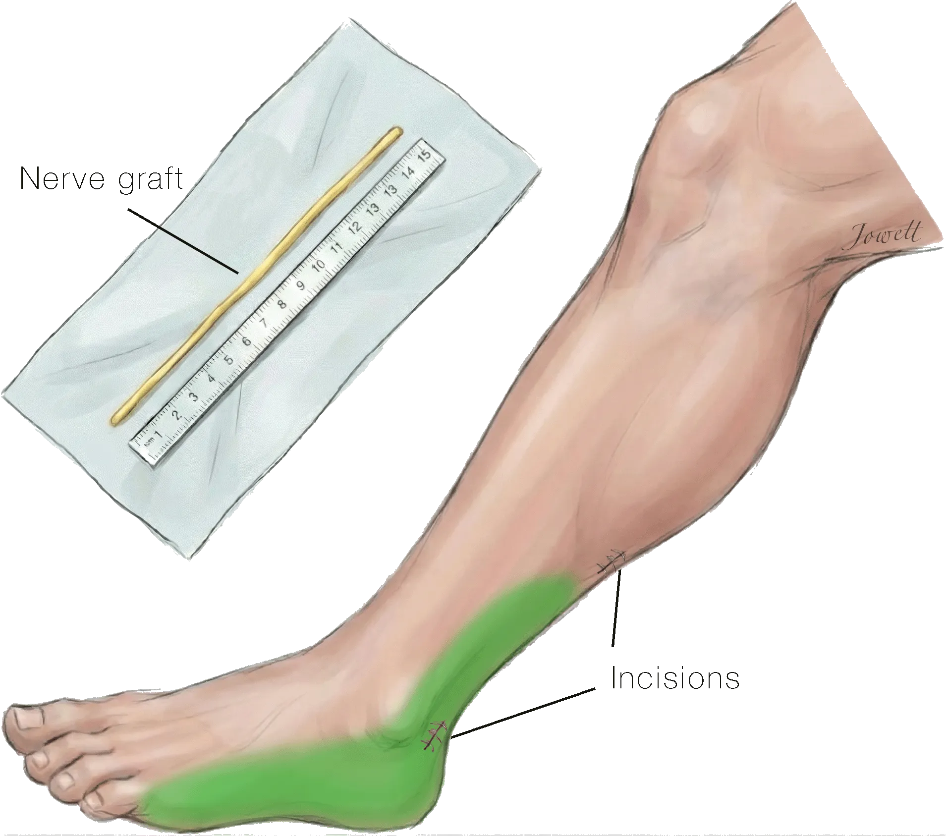
Corneal neurotization is an incredibly delicate procedure requiring collaboration between a facial reconstructive microsurgeon and a specialized ophthalmologist (cornea specialist). Two families of technique now exist. Direct transfer moves a nearby forehead nerve to the cornea. Indirect transfer uses a graft—usually sural nerve from the calf, or great auricular nerve from the neck—to bridge a longer gap.
Terzis direct transfer: Julia Terzis published the first practical series in 2009. She tunneled contralateral supraorbital and supratrochlear nerves across the forehead to the numb cornea. That proved the principle: a living sensory nerve can reawaken an anesthetic eye. The original coronal incision was large, and the method is a poor fit when both eyes are numb.
Borschel indirect sural graft: Gregory Borschel, Asim Ali, and the Toronto SickKids group then made the operation minimally invasive. A sural nerve graft is coapted to a healthy supratrochlear nerve and fanned around the limbus. That approach works in children, in bilateral disease, and when the forehead nerves on the affected side are themselves dead.
Scleral-corneal tunnel incisions: Dr. Jowett refined how the nerve enters the eye. Rather than laying fascicles only on the surface, his approach uses tiny scleral-corneal tunnels so the transferred fibers sit in the anterior stroma with less scarring and better optical clarity. He often uses the patient's own great auricular nerve, which bypasses a failed trigeminal system entirely. He argues against cadaveric acellular allografts: a living autologous graft carries Schwann cells that the cornea actually needs.
What the Evidence Shows
Corneal neurotization treats the numb eye at its source. The evidence is observational—case series, one prospective comparison, and reviews—not a randomized trial against drops. The surface benefit is still the most consistent finding in the field.
- Terzis's 2009 series of six eyes established the operation. Sensation rose from essentially none to a mean of about 278 mm on Cochet-Bonnet testing, and every eye stayed free of ulcers across a mean 16-year follow-up.
- Elbaz, Bains, Borschel, and Ali then showed that a sural graft to the supratrochlear nerve can restore feeling through small incisions. Catapano and Fung later reported 19 eyes: sensation rose from 0.8 mm to 49.7 mm, recurrent epithelial defects fell from 89% to 21%, and confocal microscopy plus magnetoencephalography proved the new nerves were real and connected to the brain.
- Woo, Daeschler, Borschel, and Ali later confirmed that the same minimally invasive operation protects against recurrent ulceration and can improve acuity when the surface is still salvageable.
- Fogagnolo's 2020 multicenter series compared direct and indirect transfer in 26 eyes. Neurotrophic keratopathy healed in every patient at a mean of 3.9 months. Sensation improved in both groups, and the two techniques were statistically equivalent at one year.
- Park's 2020 review of 54 published eyes found better sensation and vision after neurotization, with children recovering more feeling and established stromal scarring as the usual limit on acuity.
- Jowett and Pineda described great-auricular transfer through scleral-corneal tunnels and argued that the patient's own nerve, not a processed cadaver graft, is the reliable conduit. Borschel's later 10-year technique update also includes great-auricular donor transfer when the trigeminal system is gone.
- The aim is a stable, feeling surface—not a new blink. If the facial nerve is also out, eyelid weights or slings still have to close the eye. Earlier referral, before irreversible stromal scarring, gives the better sensory result and can make a later corneal transplant possible.
- Donor-nerve choice follows the map of what sensation is left. Trigeminal branches such as the supratrochlear or supraorbital nerve are preferred when they are still working, and the greater auricular nerve is used when facial sensation on that side is absent - so charting the pattern of numbness is a planning step, not a formality.
- Surgery does not replace surface care. Preservative-free lubricants, bandage contact lenses, autologous serum or plasma drops, topical nerve growth factor, punctal plugs, amniotic membrane, and partial eyelid closure may all still be needed before and after neurotization.
- When a corneal transplant is also needed, it is typically deferred about a year and until sensation and the ocular surface have been adequately restored, because a sensate, stable cornea makes the graft safer.
- Reported complications are generally low and mostly self-limiting - numbness or discomfort where a graft is harvested, eyelid swelling or scarring, and infrequent surgical problems. Serious vision-threatening complications are uncommon.
Why Choose Revitalis for Corneal Neurotization?
Dr. Nate Jowett is a global leader and innovator in corneal neurotization. The field begins with Terzis's direct transfer and the Borschel–Ali SickKids sural-graft series. His own papers added great-auricular transfer, scleral-corneal tunnels, and a critical review of which grafts actually regenerate.
Patients with complex skull-base tumors may lose both the facial nerve needed for blinking and the trigeminal nerve needed for corneal sensation. In selected cases, a coordinated plan can address both deficits by combining eyelid support with corneal neurotization. Ongoing corneal care remains essential, and surgery cannot guarantee sensation, healing, or preservation of vision.
Common questions
Questions Patients Ask
Who may benefit from corneal neurotization?
It is considered for persistent corneal anesthesia with recurrent epithelial breakdown or neurotrophic keratopathy despite appropriate medical care. The eye must be assessed for additional causes of poor healing that the nerve procedure will not correct.
Does the surgery guarantee better vision?
No. Restoring sensation can improve corneal stability and healing, but final vision depends on existing scarring, infection history, ocular-surface health, and other eye conditions.
What are the tradeoffs at the donor nerve?
Temporary or persistent numbness can occur in the donor nerve territory. Direct and indirect methods have different incision, graft, and sensory tradeoffs that should be reviewed with both reconstructive and corneal specialists.
Which nerve is used as the donor, and how is that decided?
It depends on where your facial sensation is still intact. Trigeminal branches such as the supratrochlear or supraorbital nerve are preferred when they are working, and the greater auricular nerve is used when sensation on that side of the face is absent. Mapping the pattern of numbness before surgery is therefore a genuine planning step.
Can both eyes be treated, or only one?
Indirect neurotization, which uses a nerve graft as a bridge, extends the operation to both eyes and to patients who have lost sensation on the same side of the face. Direct and indirect techniques have shown comparable healing of the ocular surface and improvement in stage, with similar overall outcomes at one year.
Will I still need drops after neurotization?
Usually yes. Surgery addresses the missing nerve supply, but supportive surface care remains important before and after - preservative-free lubricants, bandage contact lenses, autologous serum or plasma drops, topical nerve growth factor, punctal plugs, amniotic membrane, or partial eyelid closure may all still be part of the plan.
If I need a corneal transplant, can it be done at the same time?
Generally no. A transplant is typically deferred about a year and until sensation and the ocular surface have been adequately restored, because a cornea that can feel and heal makes the graft safer. Restoring sensation first is part of what makes a later transplant a reasonable option.
Answers are general and individualized at consultation.
Medical References & Evidence-Based Guidelines
- Corneal Neurotization: A Novel Solution to Neurotrophic Keratopathy. Terzis JK, Dryer MM, Bodner BI. Plast Reconstr Surg. 2009;123(1):112-120. PMID: 19116544.
- Restoration of Corneal Sensation With Regional Nerve Transfers and Nerve Grafts: A New Approach to a Difficult Problem. Elbaz U, Bains R, Zuker RM, Borschel GH, Ali A. JAMA Ophthalmol. 2014;132(11):1289-1295. PMID: 25010775.
- Corneal Neurotization from the Supratrochlear Nerve with Sural Nerve Grafts: A Minimally Invasive Approach. Bains RD, Elbaz U, Zuker RM, Ali A, Borschel GH. Plast Reconstr Surg. 2015;135(2):397e-400e. PMID: 25626824.
- Treatment of Neurotrophic Keratopathy with Minimally Invasive Corneal Neurotisation: Long-term Clinical Outcomes and Evidence of Corneal Reinnervation. Catapano J, Fung SSM, Halliday W, et al. Br J Ophthalmol. 2019;103(12):1724-1731. PMID: 30770356.
- In Vivo Confocal Microscopy Reveals Corneal Reinnervation After Treatment of Neurotrophic Keratopathy With Corneal Neurotization. Fung SSM, Catapano J, Elbaz U, Zuker RM, Borschel GH, Ali A. Cornea. 2018;37(1):109-112. PMID: 29053558.
- Corneal Neurotization Improves Ocular Surface Health in a Novel Rat Model of Neurotrophic Keratopathy and Corneal Neurotization. Catapano J, Antonyshyn K, Zhang JJ, Gordon T, Borschel GH. Invest Ophthalmol Vis Sci. 2018;59(11):4345-4354. PMID: 30193306.
- Minimally Invasive Corneal Neurotization Provides Sensory Function, Protects Against Recurrent Ulceration, and Improves Visual Acuity. Woo JH, Daeschler SC, Mireskandari K, Borschel GH, Ali A. Am J Ophthalmol. 2022;241:179-189. PMID: 35513030.
- Minimally Invasive Corneal Neurotization: 10-Year Update in Technique Including Novel Donor Transfer of the Great Auricular Nerve. Gross JN, Bhagat N, Tran K, et al. Plast Reconstr Surg. 2024;154(4):795e-798e. PMID: 38194587.
- Direct Versus Indirect Corneal Neurotization for the Treatment of Neurotrophic Keratopathy: A Multicenter Prospective Comparative Study. Fogagnolo P, Giannaccare G, Bolognesi F, et al. Am J Ophthalmol. 2020;220:203-214. PMID: 32659280.
- Corneal Neurotisation by Great Auricular Nerve Transfer and Scleral-Corneal Tunnel Incisions for Neurotrophic Keratopathy. Jowett N, Pineda Ii R. Br J Ophthalmol. 2019 Sep; 103(9):1235-1238. PMID: 30470713.
- Acellular Nerve Allografts in Corneal Neurotisation: An Inappropriate Choice. Jowett N, Pineda Ii R. Br J Ophthalmol. 2020 Feb; 104(2):149-150. PMID: 31719110.
- Seeing Through the Evidence for Corneal Neurotization. Jowett N, Pineda R. Curr Opin Otolaryngol Head Neck Surg. 2021 Aug 01; 29(4):252-258. PMID: 34074877.
- Corneal and Facial Sensory Neurotization in Trigeminal Anesthesia. Jowett N, Pineda R. Facial Plast Surg Clin North Am. 2021 Aug; 29(3):459-470. PMID: 34217450.
- Neurotrophic Keratopathy. Dua HS, Said DG, Messmer EM, et al. Prog Retin Eye Res. 2018;66:107-131. PMID: 29698813.
- Corneal Neurotization: A Review of Pathophysiology and Outcomes. Park JK, Charlson ES, Leyngold I, Kossler AL. Ophthalmic Plast Reconstr Surg. 2020;36(5):431-437. PMID: 31923091.
- Corneal Neurotization for Neurotrophic Keratopathy: Review of Surgical Techniques and Outcomes. Liu CY, Arteaga AC, Fung SE, Cortina MS, Leyngold IM, Aakalu VK. Ocul Surf. 2021;20:163-172. PMID: 33647470.
- Sural Nerve Graft for Neurotrophic Keratitis: Early Results. Weis E, Rubinov A, Al-Ghoul AR, Yau FM. Can J Ophthalmol. 2018;53(1):24-29. PMID: 29426435.
- Current Concepts in Corneal Neurotization. Ashton C, Borschel GH, Mireskandari K, et al. Am J Ophthalmol. 2026;290:101-112. PMID: 42285466.
- Insights on the Choice and Preparation of the Donor Nerve in Corneal Neurotization for Neurotrophic Keratopathy: A Narrative Review. Serra PL, Giannaccare G, Cuccu A, et al. J Clin Med. 2024;13(8). PMID: 38673540.
- Neurotrophic Keratopathy: Current Concepts, Management, and Emerging Treatments. Cheung AY, Holland EJ. Cornea. 2026;45(6):673-685. PMID: 42085714.
- Corneal Neurotization: A Meta-analysis of Outcomes and Patient Selection Factors. Swanson MA, Swanson RD, Kotha VS, et al. Ann Plast Surg. 2022;88(6):687-694. PMID: 35502965.
- Long-term outcomes of corneal neurotization for neurotrophic keratopathy: a dual center, single surgeon retrospective study. Robbins JO, Okrent AL, Grigore E, et al. Plast Reconstr Surg. 2025. PMID: 41428966.